3D Pore Network Models
![3D plot of the throat barriers (purple) and medial axis (rainbox colored) in a [0,99]x[0,99]x[0,99] voxel subvolume of the wheat using X-ray CT images](/images/3DPoreNetworkModels/preview/160s160/3Dporeimg1.jpg) 3D plot of the throat barriers (purple) and medial axis (rainbox colored) in a [0,99]x[0,99]x[0,99] voxel subvolume of the wheat using X-ray CT images
3D plot of the throat barriers (purple) and medial axis (rainbox colored) in a [0,99]x[0,99]x[0,99] voxel subvolume of the wheat using X-ray CT images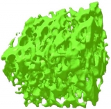 3D visualization of pore partitioning inside durum wheat grain bulk using X-ray CT tomograms
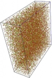 Euclidean view of the 3D medial axis of the wheat bulk
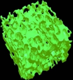 Hollow Magik Cube ~ Reconstructed intergranular airspace inside barley grain bulk using marching cube algorithm from X-ray CT images
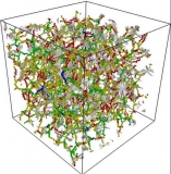 Kaleidoscope ~ Pore Network Inside Wheat Bulk
 Reconstructed bulk grain space complex surface model using X-ray CT images
Bacterial Biofilms
- AFM image of biofilm formed by YR343 field strain
- Biofilm samples stained with Calcofluor. Bacterial cells associated closely with cellulose fibres seen at high magnification
- Biofilm formed at 5 uL per hour flowrate inside a microfluidic device
- Cell clumping and arrangement of Calcofluor stained cellulose fibrils in the field isolated GM30 bacteria
- Expression of pili indicating firm adhesion of YR343 on mica substrate after 8 hours biofilm formation
- Nanoscale image of the endophytic bacteria GM30 biofilm with flagella
- GFP expressing YR343 biofilm surrounding Arabidopsis root surface
- YR343 biofilm development after 4 hours
- Field isolated bacterial strain shown by 3D reconstruction of a confocal image after 72 hours of biofilm growth
- Tapping mode AFM images of a mica surface with scattered and clustered YR 505 cells
- Pseudomonas Aeruginosa cell flocculation when stained with calcofluor
- Visualization of flagella, EPS surrounding the YR343 field strain
- High resolution confocal image of YR343 bacteria stained with calcofluor
- Nanoscale cell surface morphology of Pseudomonas Aeruginosa visualized using AFM
Biopolymer
- AFM image revealing the structure of buckwheat starch granule with alternating amorphous and crystalline zones constituting the growth ring
- Durum wheat starch granule surface
- Protein bodies (Pb) and growth rings visualized on the surface of starch granules using AFM
- Martian craters ~ undulations and pit-like shape observed on the buckwheat starch Granule Surface
- Visualization of the nanoscale non-vitreous durum wheat starch granule surface
- Error Signal mode AFM image of durum wheat starch granule surface
Cells
- AFM image of the field isolate YR343 strain with its single polar flagellum
- Populus root YR 505 strain bacterium with multiple polar flagella
- Geobacterial strain 300 revealing nano-wires
- AFM ~ Fimbriae help the field isolate YR505 strain to attach to the populus root surface
- AFM image of the isolated nanowires (~ 6 nm) from Shewanella MR1 on a mica substrate
- SEM image of rod shape field isolate GM01 strain ~200 nm~
- Nanowires observed surrounding the cell surface of a field isolate grown in Differential minimal media with the electron donor 10 mM Lactate
- Anti microbial peptide BP100 effect on GM30 strain - cell perturbation and disruption
- Nanoscale visualization of the endophyte GM30 strain cell surface structure using AFM
- TEM image of GM30 strain with its single polar flagellum
- AFM ~ YR343 strain with fimbriae
- TEM image of YR343 strain with its single polar flagellum
- AFM ~ E.Coli DH5 alpha with flagella
Chips and Nanotools
- Functionalized silicon cantilever tip for Force Spectroscopy
- Interdigitated gold electrode array for sensor platforms
- Probing bacterial interaction forces with functionalized cantilever
- Cross-sectional view of the developed gas sensor sensor (i) Gold interdigitated electrode PCB array, (ii) PABA polymer, (iii) Nafion, (iv) O-ring, (v) Gas permeable membrane.
Chromosomes
- AFM image of buckwheat genomic DNA on mica
- Error Signal Mode non-Contact AFM Image of Fagopyrum Esculentum (common buckwheat) chromosomes in Air
- Complete Set of Common BuckWheat Chromosomes (AFM topographic mode)
- Confocal image of DAPI stained Agave Americana Marginata chromosomes
- Great Sand Dunes ~ 3D surface reconstructed atomic scale images of buckwheat chromosomes Scale 180 nm
- AFM image of polyaniline templated on stretched genomic DNA from buckwheat
- Error Signal Mode AFM Image of Common Buckwheat chromosomes in Air
- Agave Tequilana chromsomes spread on cover glass as observed under Confocal Microscope
- Tartary buckwheat chromosome spread on cover glass
- AFM image of spread of Fagopyrum Tartaricum chromosome set
- Fagopyrum Tartaricum chromosome spread on glass slide
Grain Kernels
Microfluidics
- Serpentine shape microfluidic device
- Concentration gradient observed in the serpentine microfluidic channel
- Multiple output gradient generating microfluidic device
- Gradient generating microfluidic device for bacterial chemotaxis ~ inlet
- Gradient generating microfluidic device for bacterial chemotaxis ~ mixing
- Close up of the serpentine mixing channel
- Gradient generating microfluidic device for bacterial chemotaxis ~ Feeder
- Integrated nanoporous microfluidic device for characterizing single cell motility
- Nanoporous microfluidic device for bacterial chemotaxis
- Nanoporous fluidic device
- Nanopores revealed in the microfluidic channel device
Nanoparticles
- Platinum Flowers ~ Nanoparticle lattice formation observed using topopgraphic AFM image
- AFM image of aggregated silica nanoparticle
- AFM topography image of rod shaped starch nanocrystals from a dried water suspension
- Aggregated clumps of biopolymer remnants
- Nanoparticles from platinum colloid
- Lattice formation by nanoparticles from dried platinum nanocolloid
Plant-Microbe Interactions
- GM30 endophytic colonization inside plant root tissue
- Live-dead staining of bacteria on root surface of Arabidopsis
- YR343-Biofilm formation along the root hair zone of Poplar tree root
- Dried Arabidopsis root colonized with GM30 bacterial strain
- Biofilm colonization along the plant cell wall ~ Xylem is visible in the confocal image
- Live bacteria cells surrounding the root hair zone
- Biofilms of Rhizobium sp developed on the roots of A. thaliana plantlets grown using Confocal
- YR343 colonization of the surface of the Poplar root hair zone
- Colocalization of YR343 bacterial strain expressing the GFP after propidium-iodide labelling
- Evidence of YR343 biofilm formed on Poplar tree roots grown
- Biofilm between root crevices
- SEM micrograph of Poplar tree root colonized by YR 343 bacterial strain
- Root surface colonized with bacteria
